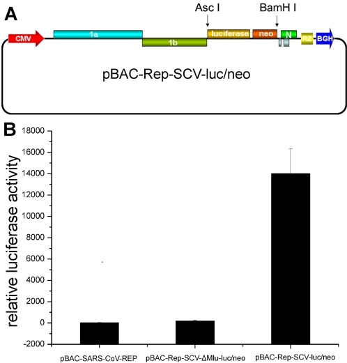
Figure 3. Structure and activity assay of the reporter replicon construct pBAC-Rep-SCV-luc/neo.
(A) Schematic structure of the replicon. The coding sequence of luciferase-neomycin fusion under the control of M gene TRS was inserted into the basic replicon construct pBAC-SARS-CoV-REP between AscI and BamHI sites (For details, see the Materials and Methods). (B) Luciferase assays of the reporter replicons. 2×10⁵ BHK21 cells were transfected with the three kinds of replicon plasmids (0.4 µg each), respectively, and pRL-TK plasmid (0.1 µg) as an internal control. The luciferase activity assays were performed 24 h post transfection. Error bars represent standard deviations of the mean of three experiments.

