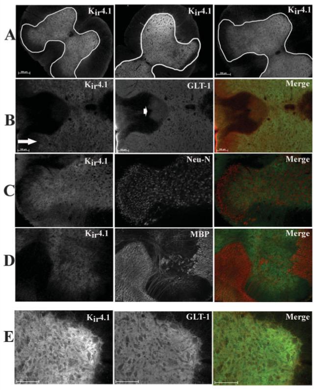
Fig. 10.

Immunohistochemistry demonstrated Kir4.1 is prominently expressed in the gray matter astrocytes in spinal cord in vivo. All images were from fixed, 50-μm sections from the cervical, lumbar, and thoracic regions. A: Kir 4.1 is expressed throughout the gray matter in each spinal cord section (scale bar 250 μm). B: Kir4.1 and GLT-1 (an astrocyte-specific glutamate transporter) largely overlap in the gray matter, however, GLT-1-positive cells in the apex of the dorsal horn (arrow) and astrocytes in the dorsal corticospinal tracks (arrowhead) that demonstrated very little Kir4.1 immunoreactivity. C: Kir4.1 and Neu-N (a neuronal marker) did not demonstrate similar staining patterns in the spinal cord slice. D: Kir4.1 and MBP (myelin basic protein) an oligodendrocyte-specific marker, demonstrate very little overlap in either the white matter or gray matter in vivo. E: Higher-magnification image of Kir4.1 and GLT-1 demonstrate overlap of the two proteins in the spinal cord slice. Scale bar = 100 μm in B,E.
