
Fig. 2.
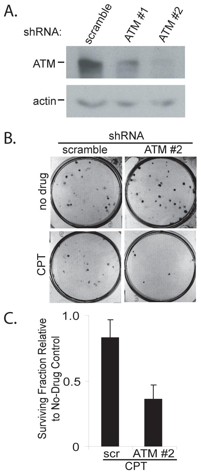
ATM depletion sensitizes LOX cells to camptothecin. A. LOX melanoma cells were infected with lentivirus expressing the indicated shRNAs and cells were harvested 4 days later. ATM knockdown was analyzed by Western blot, with actin serving as a loading control. B. LOX cells were infected with scramble #1 or ATM #2 shRNAs and seeded in 6-well plates 3 days later at a density of 100 cells per well. After twelve hours, the cells were treated with 50 nM camptothecin in DMSO (CPT) or DMSO alone (no drug). The medium was replaced 24 hours later and the cells were grown for an additional 6 days. Cells were fixed and stained with crystal violet to assess colony number, and images of representative wells are shown. C. The graph shows the fraction of LOX colonies surviving camptothecin treatment relative to the corresponding DMSO controls. The data are derived from three independent experiments. Error bars indicate the standard deviation, and the difference between the samples is statistically significant (p = 0.01).