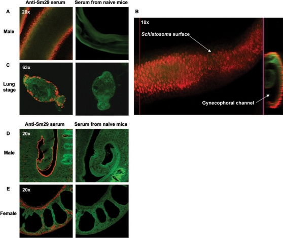
Figure 1. Immunolocalization of Sm29 antigen on male and female adult worm and lung-stage schistosomula of S. mansoni.
Polyclonal anti-Sm29 antibodies, serum from mice that received Freund́s adjuvant as negative control, and Cy5-conjugated anti-mice IgG were used. Actin was visualized by falloidin-Alexa fluor 488. The parasites were fixed in Omnifix II and used to whole-mount or section immunolocalization. (A and B) Whole-mount immunolocalization of Sm29 antigen on the surface (outer tegument) of male adult worm and (C) lung-stage schistosomula of S. mansoni. (D) Immunolocalization of Sm29 on the surface (outer tegument) of male adult worm, and (E) on the surface (outer tegument) and in some internal tissues on the female adult worm using deparaffinized sections of the parasites. Localization of Sm29 is identified by the orange color and actin filaments by the green color.

