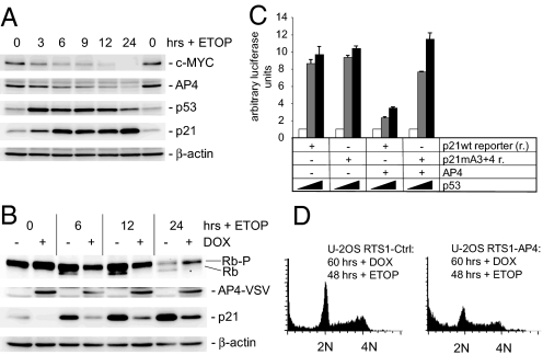
Fig. 4.
Role of AP4 in the DNA damage response. (A) Effect of DNA damage on AP4 expression. MCF-7 cells were treated with etoposide (ETOP, 20 μg/ml) and cell extracts were obtained at the indicated time points. Expression of the indicated proteins was determined by immunoblotting. (B) Ectopic AP4-VSV was induced in U-2OS cells for 12 h by addition of DOX (100 ng/ml). Then ETOP (20 μg/ml) was added for the indicated periods. Expression of the differentially phosphorylated retinoblastoma protein (Rb-P/Rb), AP4-VSV, p21 or β-actin was detected by immunoblotting. (C) p21 reporter activity was determined in H1299 cells transfected with the indicated plasmids. Increasing p53 expression was achieved by transfection of 0, 50 or 200 ng of plasmids (indicated as  ). Shown are the median expression values and standard errors of two independent transfection experiments. p21 mA3 + 4: see Fig. 3A. (D) AP4 was induced by DOX for 12 h before treatment of cells with ETOP (20 μg/ml) for 48 h. Then cells were analyzed by flow cytometry. Depicted are exemplary histograms representing 10,000 cells. 2N: cells in G1, 4N: cells in G2/M.

