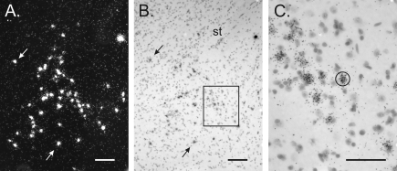
Figure 1.
In situ hybridization for VP mRNA in a Bcl-2-OE male. A and B, Dark- and bright-field views, respectively, of a section through the BNST. Counts of VP cells were made under dark-field microscopy. Arrows point to the same cells in A and B. A magnocellular VP neuron, easily distinguishable from the parvocellular BNST neurons of interest, can also be seen in the upper right corner. St, Stria terminalis. C, Higher-magnification view of the boxed area in B showing sliver grains over individual VP-positive neurons. A standard circle, as shown here, was centered over each cell for automated grain counting. Scale bars, 100 μm (A and B) and 50 μm (C).

