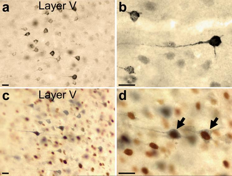
Figure 2.
Representative micrographs of deep layers of the mPFC showing FG-filled neurons (DAB-Ni) at (a) 20X and (b) 60X, and neurons dually immunolabeled for Fos (DAB) and FG (Vector SG) at (c) 20X and (d) 60X. Dually immunolabeled neurons (arrows) have an amber nucleus (Fos-immunoreactive cell) surrounded by blue cytoplasm (FG-immunoreactive neuron). Scale bars: a-d, 25 μm.

