Figure 2.
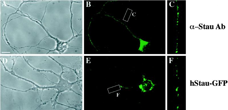
Expressed human Staufen-GFP and endogenous rat Staufen protein show a comparable punctate, dendritic expression pattern in hippocampal neurons. Adult rat hippocampal neurons were either fixed and labeled with anti-hStau antibodies (A–C) or transiently transfected with hStau-GFP and fixed, and green fluorescence was recorded (D–F). (A–C) Phase-contrast and hStau immunofluorescence of a representative neuron. C is an enlarged section of the white box in B. (D–F) Phase-contrast and hStau-GFP fluorescence of a representative neuron. F is an enlarged section of the white box in E. The small precipitate seen in D is due to CaPi transfection. Note that two different types of fluorescent granules were routinely observed (E): large granules around the nuclear membrane and smaller granules in the periphery of the neurons. Bar, 10 μm.

