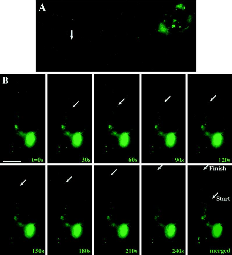
Figure 4.
Individual hStau-GFP particles are transported into dendrites of living hippocampal neurons. Hippocampal neurons on glass coverslips were transfected with hStau-GFP as described in Figure 2, and individual motilities of green fluorescent granules were observed by time-lapse video microscopy. (A) Corresponding figure to Video 1; (B) corresponding figure to Video 2. In both videos as well as in B, moving particles are labeled by an arrow and were followed during its anterograde transport into the dendrite. The individual video frames were more highly integrated compared with Fig. 2 to detect individual granules in processes. Elapsed time is indicated in seconds in the lower right of each video frame in B. The last panel in B shows a merged picture of all frames depicting both the Brownian movement of most particles as well as the discontinuous, directed motility of the chosen granule. Bar, 10 μm.

