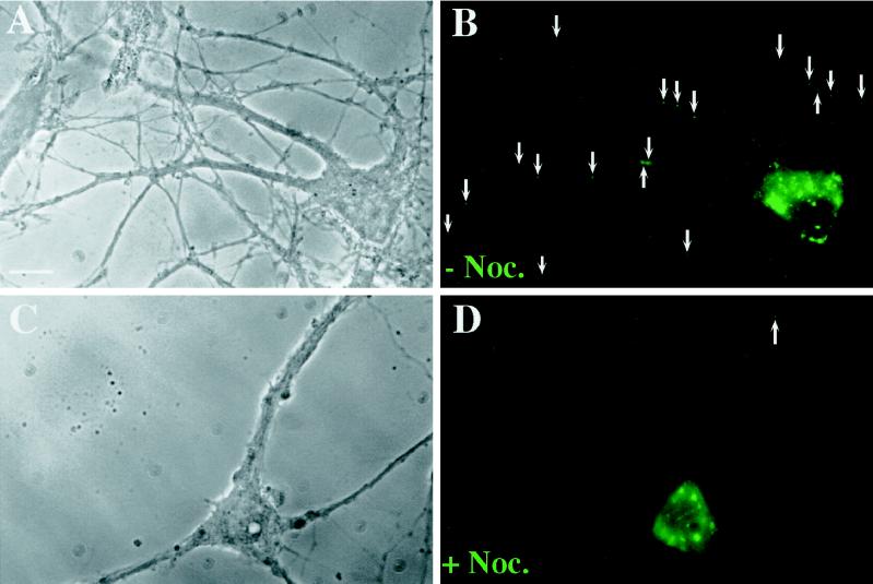
Figure 6.
Nocodazole treatment of hippocampal neurons results in a diminished number of Stau-GFP particles in distal dendrites. Hippocampal neurons were transiently transfected as described in Figure 2 and the following day were treated with nocodazole (20 μM) for 3.5 h or mock treated. Cells were fixed, and fluorescence was recorded. (A and B) A representative cell is shown containing 19 Stau-GFP granules (arrows) in distal dendrites (>12 μm apart from the cell body). (C and D) A typical nocodazole-treated cell with a granular expression pattern is shown containing one granule (arrow) in distal dendrites. Bar, 10 μm.

