
Figure 1. Time course of [3H] biotin uptake in MDCK-MDR1 cells.
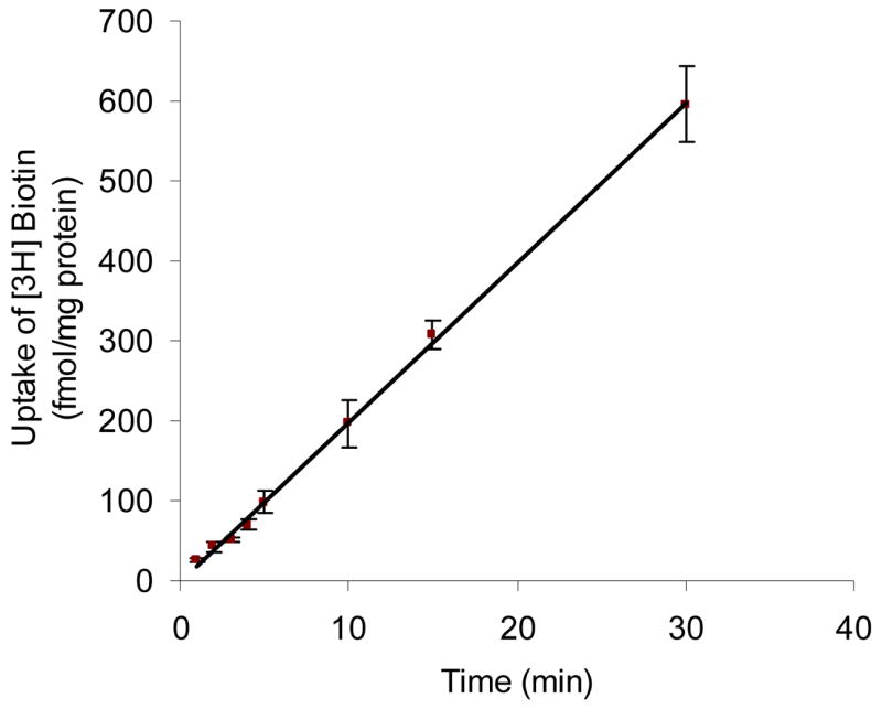
Uptake of [3H] biotin (10 nM) was measured in DPBS buffer (pH 7.4) at 37 °C. Data are shown as mean ± SD, n = 3–6. The linear equation is represented as: y = 20.0 × − 1.57 (r2 = 0.999).
Official websites use .gov
A
.gov website belongs to an official
government organization in the United States.
Secure .gov websites use HTTPS
A lock (
) or https:// means you've safely
connected to the .gov website. Share sensitive
information only on official, secure websites.
Uptake of [3H] biotin (10 nM) was measured in DPBS buffer (pH 7.4) at 37 °C. Data are shown as mean ± SD, n = 3–6. The linear equation is represented as: y = 20.0 × − 1.57 (r2 = 0.999).