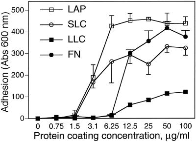
Figure 1.
A549 cell adhesion to LAP, SLC, LLC, and FN. (A) Wells were coated with solutions of LAP, SLC, LLC, and FN, at various concentrations as shown, and blocked with BSA. A549 cells were allowed to adhere to the coated wells for 90 min, and nonadherent cells were washed off. Absorption at 600 nm indicates the amount of stain associated with adherent cells and is proportional to cells bound.

