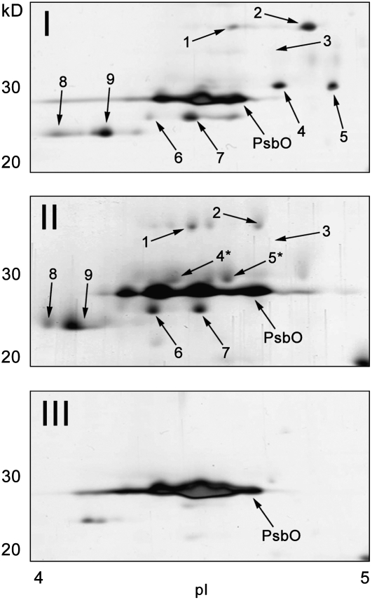
Figure 5.
Two-Dimensional Separation of LHCII Polypeptides in the Gel Filtration Fractions I to III.
Fractions I to III isolated from a State 2–locked sample were subjected to 2-DE. Shown are the regions between 20 and 40 kD and pH 4.0 and 5.0 on the silver-stained 2-DE gels. Each numbered spot was identified by MS/MS analysis (see Supplemental Table 2 online) after in-gel trypsin digestion. Asterisks indicate putatively phosphorylated polypeptides (see corresponding spots 13 and 14 in Figure 8).

