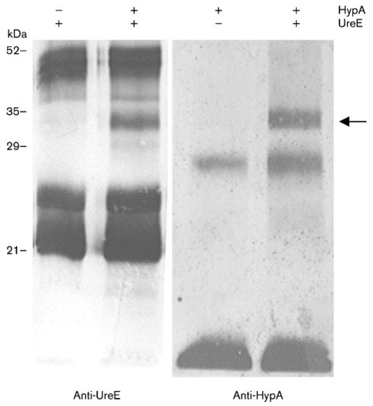
Fig. 2.
Immunoblotting analysis of cross-linked products arising from a mixture of HypA and UreE. The cross-linking was carried out in presence (+) or absence (−) of HypA or UreE, as indicated above the panels. The membrane was probed with either anti-HypA or anti-UreE antiserum, as shown below each panel. The position of the HypA–UreE adduct is shown with the arrow. Sizes of molecular mass standards are indicated on the left.

