
Figure 1.
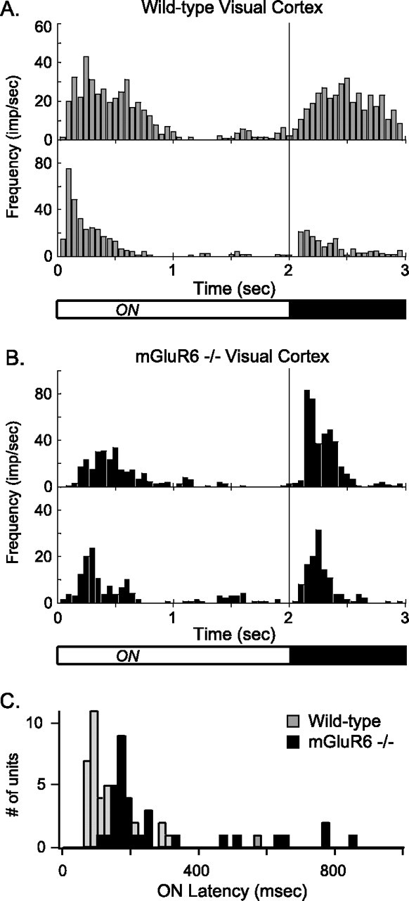
Neurons in the visual cortex of mGluR6 homozygous null mice respond to the onset of light stimuli with a long-latency response. Multiunit, extracellular electrode recordings of responses to 2 s, full-field light presentations were made from the visual cortices of WT and mGluR6-null mice anesthetized with urethane. A, Peristimulus time histograms (PSTHs) of two recordings from neurons in WT visual cortex. B, PSTHs of two recordings from neurons in visual cortex of the mGluR6 homozygous null mouse. Bin width, 50 ms. The light/dark bar below the histograms represents the light stimulus, which originated from a computer monitor. C, Summary of visual cortical neuron latencies to peak frequency after light onset. Light bars show WT latencies; black bars show mGluR6-null latencies. Neurons in the mGluR6 null have a longer latency on average compared with that of WT. Frequency is in impulses (spikes) per second (imp/sec).