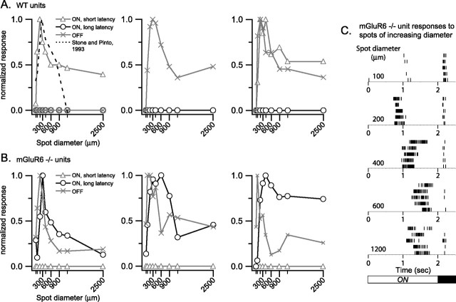
Figure 5.
The long-latency ON response of ganglion cell neurons in the mGluR6 homozygous null retina has center-surround characteristics. Receptive field organization of individual neurons was determined using nine light spots of increasing diameter presented three or five times each. Total spike counts for windows of time representing short (open triangles)- and long (open circles)-latency ON responses and OFF responses (X symbols) were normalized to the maximum count for each neuron. A, Examples of WT responses indicating typical organization of the center and antagonistic surround for an ON, OFF, and ON–OFF neuron are shown. The antagonistic surround depresses the spiking response during the presentation of the larger diameter spots. The previously published data of Stone and Pinto (1993) (dashed line) from intracellular recordings of whole-mounted mouse retina are presented in the left example. B, Examples of receptive field organization based on responses recorded from mGluR6-null retinas are shown. No short-latency ON responses were observed. Of the nine spot sizes presented, the optimal size for the long-latency response tended to be larger than that for the OFF responses in the same neurons and larger than the optimal WT spot size. Nonetheless, an antagonistic surround was observed, suppressing the responses to larger spot sizes. C, Raster plots of the spike responses to multiple presentations of spots of increasing diameter are displayed for the neuron from an mGluR6-null retina that is graphed on the right in B. The increase in latency associated with increasing spot diameter is attributable to increased brightness caused by scatter in the center as the spot size is made larger as well as activation of larger amounts of the center area.

