
FIGURE 1. Proteins in the analysis.
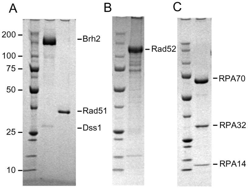
SDS polyacrylamide gels with approximately 15 pmol each of the indicated proteins run along side a ladder of Precision Protein standards (BioRad Laboratories) were stained with Comassie blue. (A) MBP-Brh2/His-Dss1 and Rad51 (B) MBP-Rad52 (C) RPA heterotrimer.