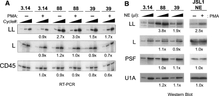
FIGURE 3.
HnRNP LL is up-regulated in clones 39 and 88 as well as upon PMA stimulation of WT cells. (A) RT–PCR analysis of hnRNP LL mRNA at 20, 24, and 28 cycles, hnRNP L mRNA at 12, 16, and 20 cycles, or 16, 20, and 24 cycles for CD45 mRNA. Fold increase of signal relative to 3.14 resting cells shown is the average difference quantitated for at least the two lowest cycle points. Constitutive exons 8–10 from the endogenous CD45 gene are used as a control. (B) Western blot analysis of hnRNP LL, L, and PSF in nuclear extracts (NE) from 3.14 cells and clones grown under resting conditions, as well as from parental JSL1 cells grown under resting (−PMA) or activated (+PMA) conditions. All extracts were normalized for total protein level prior to loading and loaded at 5 or 15 μg of total protein per lane. Expression of LL, L, PSF, or U1A (loading control) were quantitated by densitometry at both loading points and averaged to give number shown.

