FIGURE 2.
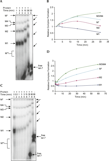
let-7 miRNPs form in different ways. (A) Time-course analysis of miRNP assembly. Gel retardation samples were withdrawn at different durations of time and loaded into a running native gel. The differential mobility of the complexes is due to time differences between each loading. Lane 6 corresponds to the free label loaded at the same time as lane 5. The shifted positions of complexes are also shown by slanted arrows on the right. In lanes 1–3, the free probe ran off the gel because of earlier loading. (B) Amounts of complexes in A at different time intervals compared to the beginning of the reaction (1 min). (C) A gel retardation reaction was chased with an excess (20-fold) of unlabeled let-7 miRNA after the first minute of incubation. At indicated time intervals, the samples were withdrawn and analyzed as described in A. (D) Amounts of complexes in C at different time intervals compared to the beginning of the pulse chase (0.5 min after addition of unlabeled let-7 miRNA).

