
Fig. 3. Del-1 gene transfer enhances vascular density.
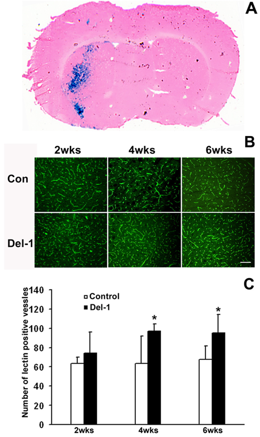
A. Distribution of X-gal positive staining (blue color) at 5 days following the injection of 5.6 × 10¹⁰ particles of AAV-lacZ. The brain section was counter-stained with H&E. B. Representative images of immunofluorescent staining of lectin positive blood vessels demonstrate that AAV-Del-1 transduction increased vascular density in the needle tract region compared to AAV-lacZ transduction. Size bar = 50µm. C. Quantification of microvessel numbers. *, p< 0.05 vs AAV-lacZ, n=8 for each group. The data are representative of 3 separate experiments.