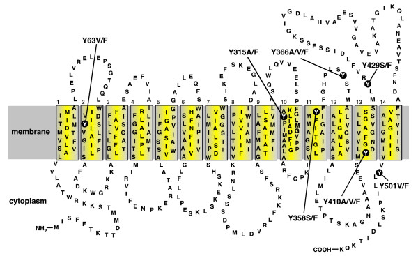
Figure 1.
Secondary structure model of the QacA multidrug transport protein. Topology of the QacA multidrug efflux protein based on hydropathy predictions and solvent accessibility analyses [6,7,10]. The membrane is depicted as a grey shaded band and the 14 TMS of QacA are shown in boxes numbered 1–14. The locations of the seven tyrosine residues and their amino acid substitutions within QacA are shown.

