
Figure 1.
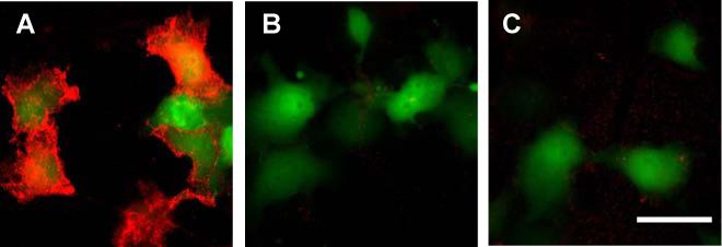
A two-step labeling of a cell surface HaloTag protein in living cells with QDs. COS7 cells expressing membrane HaloTag protein (mHTP) and EGFP as a transfection marker are incubated with (A) or without (B) HaloTag ligand 1 for 30 min in 37 °C; (C) COS7 cell expressing only EGFP are incubated with HaloTag ligand 1 for 30 min in 37 °C. After washing, these cells are then labeled with 5 nM of sQD for 30 min in room temperature before imaging. QD signal is acquired with 100 ms exposure. Shown images are overlaid frames of QD (red) and EGFP (green) fluorescence of the same region. Scale bar, 50 μm.