Figure 3.
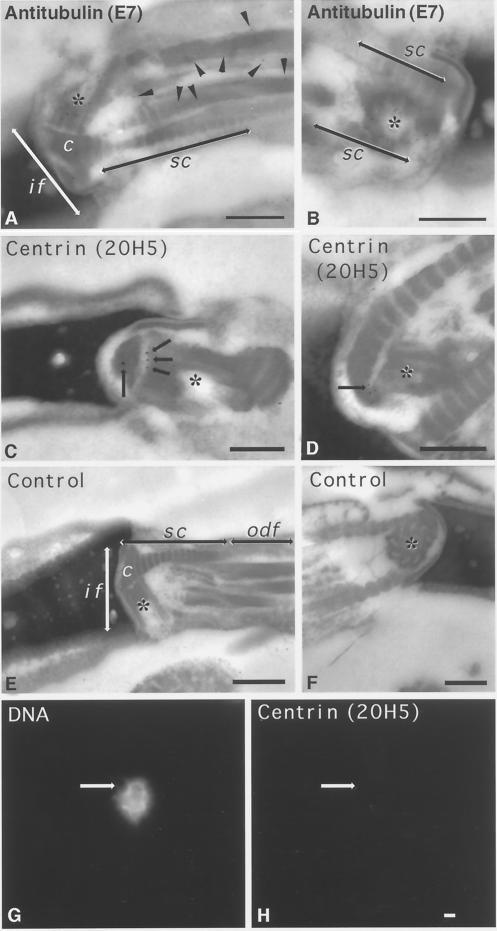
Ultrastructural detection of centrin in the bovine mature sperm centrosome and its sensitivity to CSF-arrested cell-free extract. (A and B) Immunogold labeling of mature bovine spermatozoa with anti-β-tubulin antibody, demonstrating extensive immunolabeling of the sperm proximal centriole (asterisks) and outer microtubule doublets of the sperm axoneme (A, arrowheads). (C and D) Immunogold labeling of mature bovine sperm with anti-centrin antibody 20H5. In C, a longitudinal section of proximal centriole in the sperm tail connecting piece is observed, with centrin localized to its capitulum-attached end (arrows). (D) Oblique sections of the proximal centriole demonstrating centrin detection in an area of the centriole adjacent to the striated columns of the connecting piece. (E and F) Control bovine spermatozoa immunolabeled with colloidal gold–conjugated secondary antibody only. No labeling is observed in the connecting piece structures (E, longitudinal section of the centriole) or in the proximal centriole (F, cross-section). (G and H) Human permeabilized spermatozoa treated sequentially with 5 mM DTT and CSF-arrested cell-free extract and then immunostained with the 20H5 centrin antibody. The primed sperm has begun to decondense in the presence of egg extract (G), but 20H5 centrin is no longer detected at the base of the sperm head (H, arrow). if, implantation fossa; c, capitulum; sc, striated columns; odf, outer dense fibers; asterisks, proximal centriole. Bars in A, B, D, E, and F, 0.2 μm; bar in C, 0.5 μm; bar in H, 1 μm.

