Figure 5.
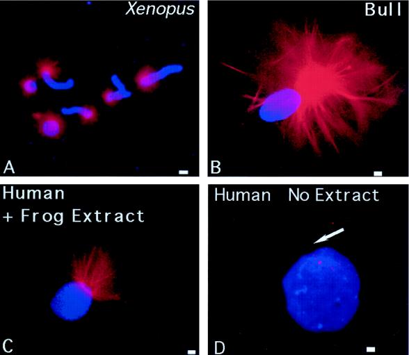
Microtubule assembly in vitro nucleated by X. laevis, human, and bovine sperm. (A) Lysolecithin-permeabilized Xenopus sperm incubated in CSF-arrested cell-free extract containing 0.08 mg/ml rhodamine-conjugated bovine brain tubulin. Microtubule assembly (red) is radially symmetric and tightly focused at the sperm centrosome (blue, DNA). Bull (B) and human (C) sperm (blue), exposed to 5 μM ionomycin and primed with 5 mM DTT, also demonstrate assembly of microtubules in vitro (red) from the centrosomal region after 40–60 min of incubation in CSF-arrested cell-free extract. No free asters or assembled microtubules are present in the background, suggesting that microtubule nucleation, as opposed to microtubule capture, has occurred. Primed human sperm (blue) that was not exposed to CSF extract did not nucleate microtubules when exposed to rhodamine-conjugated bovine brain tubulin in Pipes buffer alone (D, red; arrow points to sperm axoneme). Bar in A, 30 μm; bars in B, C, and D, 1 μm.

