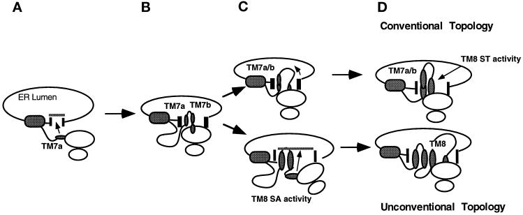
Figure 10.
Schematic model for generating topological heterogeneity in the ER membrane. Depicted are translocation events and topological intermediates proposed to participate in MDR1-Pgp topogenesis. After translocation reinitiation by TM7a (A), TM7b and TM8 emerge from the ribosome into the translocation channel (B) where the nascent chain faces two topological fates. In one cohort of chains TM7b fails to terminate translocation (C), and the chain continues to move into the ER lumen until translocation is terminated by ST activity of TM8. In the second cohort of chains, translocation is terminated by TM7b and subsequently reinitiated by the C-trans (type II) signal sequence activity of TM8. Large and small shaded ovals represent the Pgp N-terminal hydrophobic domain and individual TM segments, respectively. Translocation machinery is indicated by vertical bars. The diagram is meant only to describe the temporal sequence of specific translocation events. The precise nature of interactions among the ribosome, translocon, TM segments, and lipid bilayer is unknown.

