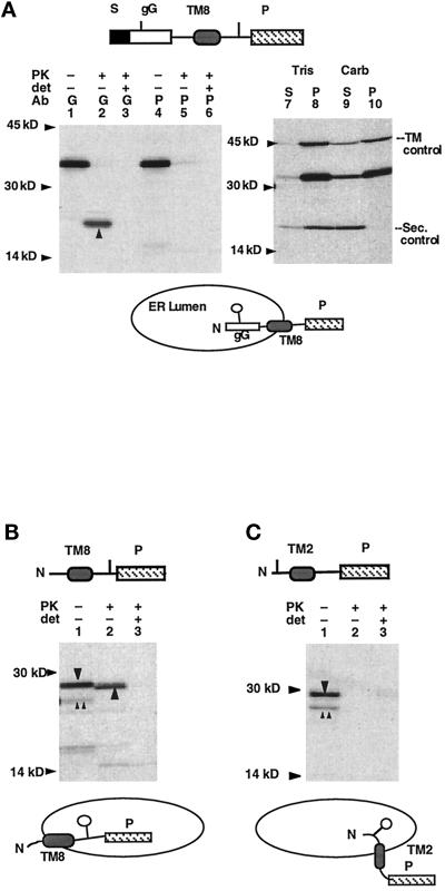
Figure 3.
TM8 topology is dependent on its mode of presentation to the ER membrane. (A) Topology of polypeptides generated from plasmid S.gG.TM8.P was determined as in Figure 2. Upward arrow (lane 2) indicates the globin-protected fragment generated by PK digestion, demonstrating a type I topology for these chains (diagrammed beneath the autoradiogram). (B and C) PK digestion of polypeptides generated from plasmids TM8.P and TM2.P. Upward double arrows and downward arrow indicate full-length unglycosylated and glycosylated chains, respectively (lane 1). Upward arrow indicates PK-protected fragment (lane 2). Potential glycosylation sites are indicated in by short vertical lines, and used glycosylation sites are indicated below by the open circles.

