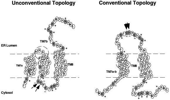
Figure 5.
Conventional and unconventional topologies of TM7a/b-TM8 in the ER membrane. Residues within predicted TM segments are indicated. Charged residues within flanking regions are shaded. Boundaries of TM segments are estimated based on the lowest free energy of transfer for partitioning the helix into the lipid bilayer and do not take into account potential interactions with adjacent helices that may shift these boundaries. Dashed circle indicates position of peptide connecting TM7a and TM7b. Arrows indicate positions of residues Asp-743 and Asp-744.

