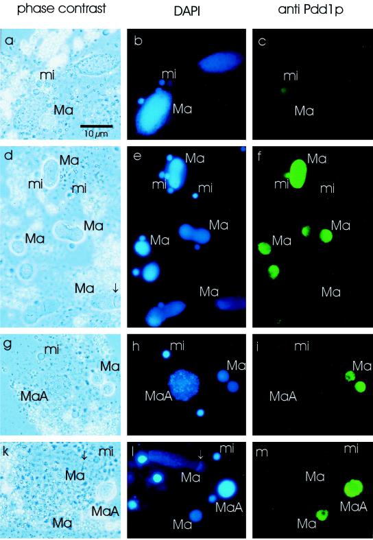
Figure 3.
Immunofluorescence analysis of vegetatively growing, starved, and exconjugant cells. Vegetative growing cells (a–c), starved cells (2 d without feeding, d–f), and exconjugant cells during polytene chromosome stage (g–i) and at a later stage (k–m) were fixed and incubated with the Pdd1p antiserum (secondary anti-rabbit Cy2-labeled antibody), and the DNA was stained with DAPI. (a, d, g, k) Phase-contrast microscopy, (b, e, h, l) DAPI stain (UV light), (c, f, i, m) anti-Pdd1p stain (blue light). Ma, macronucleus; MaA, macronuclear anlagen; mi, micronucleus. The replication band is labeled by an arrow. Bar, 10 μm.

