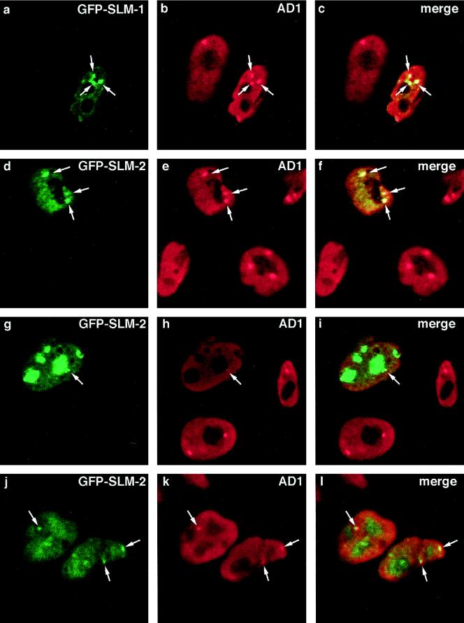
Figure 2.
GFP-SLM-1 and GFP-SLM-2 colocalize with Sam68 in nuclear bodies. GFP-SLM-1 (a–c) and GFP-SLM-2 (d–l) were transfected individually in HeLa cells, and the cells were fixed, permeabilized, and immunostained with anti-Sam68 AD1 antibody followed by a rhodamine-conjugated goat anti-rabbit secondary antibody (b, e, h, and k). Colocalization was determined by confocal microscopy, and the merged images are shown on the right (c, f, i, and l). The arrows point to nuclear bodies that colocalize. Three major localization patterns were observed with GFP-SLM-2: extranucleolar staining (d), accumulation in nucleoli (g), and diffuse staining in both nucleoplasm and nucleoli (j).

