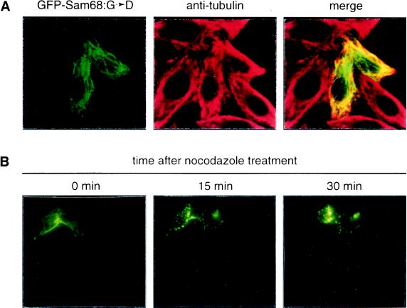
Figure 6.
Sam68:G→D and Sam68:I→N associate with microtubules. (A) HeLa cells transfected with GFP-Sam68:G→D were fixed, permeabilized, and immunostained with anti-tubulin antibody followed by a rhodamine-conjugated goat anti-mouse secondary antibody and then analyzed by confocal microscopy. Colocalization of Sam68 fibers (green) with microtubule fibers (red) resulted in yellow color when the confocal images of GFP-Sam68:G→D and anti-tubulin immunostaining were merged. (B) HeLa cells transfected with GFP-Sam68:G→D were incubated with 40 ng/ml nocodazole, and cells with fibrous phenotype were photographed live before (0 min) and 15 or 30 min after the addition of nocodazole.

