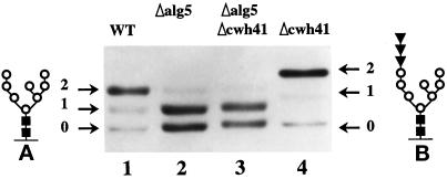
Figure 2.
N-glycosylation of Wbp1p. Total cell extracts from strains LCY21 (lane 1), LCY22 (lane2), LCY23 (lane 3), and LCY24 (lane 4) were analyzed by 8% SDS/PAGE and Western blot. Arrows indicate the number of glycosylation sites used. (A and B) Structure of the oligosaccharides, N-acetylglucosamine (closed squares), mannose (open circles), and glucose (closed inverted triangles).

