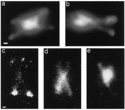
Figure 7.
(a and b) Two examples of DAPI-stained, arrested top2 cells. Chromatin fibers are stretched crosswise out of the nucleus. Bar, 1 μm. (c–e) Immunofluorescence micrographs of an arrested top2 cell at restrictive temperature. Spindle pole bodies (c) and spindles (d) were stained with specific antibodies (for details see MATERIALS AND METHODS). The nucleus is visualized by DAPI staining (e). Four spindle pole bodies (c) and two crossed meiosis II spindles (d) are visible in a cell that contains only one nucleus, of which DNA fibers are pulled out (e). Bar, 1 μm.

