Figure 10.
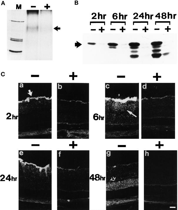
In vivo effects of SR1 on exogenous FGF2 binding and internalization in normal retina. (A) Cross-link of retinal extract with injected 125I-FGF2 in the absence (+) and presence (−) of an excess of SR1. A specific band was detected at 160 kDa (black arrow). M, molecular mass markers (200, 92, and 69 kDa). (B) Internalization and processing at 2, 6, 24, and 48 h after injection of 125I-FGF2 injected in the absence (−) and presence (+) of an excess of hrSR1. The black arrow shows intact 125I-FGF2. (C) Dark-field micrographs from F344 albino rats injected intraviteally with 125I-FGF2 in the absence (−) and presence (+) of an excess of hrSR1 and observed at 2, 6, 24, and 48 h after injection. At 2 h, labeling appeared in the ganglion cell layer (large white arrow) and then diffused in the inner nuclear (thin white arrow) and plexiform layers at 6 h. At 48 h, labeling was mainly present in the ONL (open white arrow). Comparable results were obtained in three independent experiments.

