Figure 3.
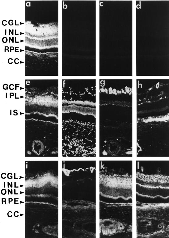
Immunohistochemical distribution of SR1 in retina. Fixed frozen rat retina were permeabilized with Triton X-100 (except in a). The slides were incubated with antibodies using a double-labeling technique (except in a–d and h) as described in MATERIALS AND METHODS. Retina sections (8 μm) were incubated with anti-SR1 antibody (1:250; a, c, e, i, and k), anti-neurofilament 68-kDa antibody (g), anti-factor VIII antibody (h), anti-GFAP antibody (j), and anti-synaptophysin antibody (l), and cell nuclei were stained with DAPI (f). Checks on anti-SR1 labeling specificity: incubation of the retina sections with 1) the control preimmune serum (b), 2) the anti-SR1 antibody preadsorbed with the cognate antigen peptide (c), and 3) primary antibody omitted (d) all gave no labeling. IPL, inner plexiform layer; OPL, outer plexiform layer; ONL, outer nuclear layer; IS, inner segment; RPE, retinal pigmented epithelium; CC, choroid; GCF, ganglion cell fiber. Bar, 55 μm.

