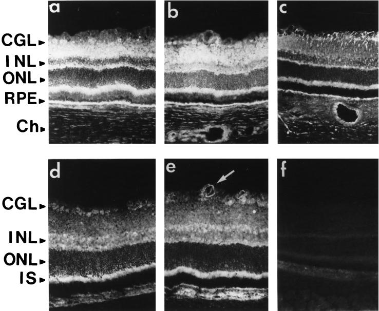
Figure 4.
Comparison of SR1 immunoreactivity with FGFR1, FGF1, and FGF2 immunoreactivities in retina. Sections of adult rat retina fixed with 4% paraformaldehyde and permeabilized with Triton X-100 (except in a) were incubated with antibodies against SR1 (1:250; a and b), the carboxyl terminus of FGFR1 (1:200; c), FGF1 (1:100; d), and FGF2 (1:100; e). The white arrow shows FGF2 staining in vascular endothelial cells. GCL, ganglion cell layer; INL, inner nuclear layer; ONL, outer nuclear layer; IS, inner segment; RPE, retinal pigmented epithelium; Ch, choroid. Bar, 70 μm.

