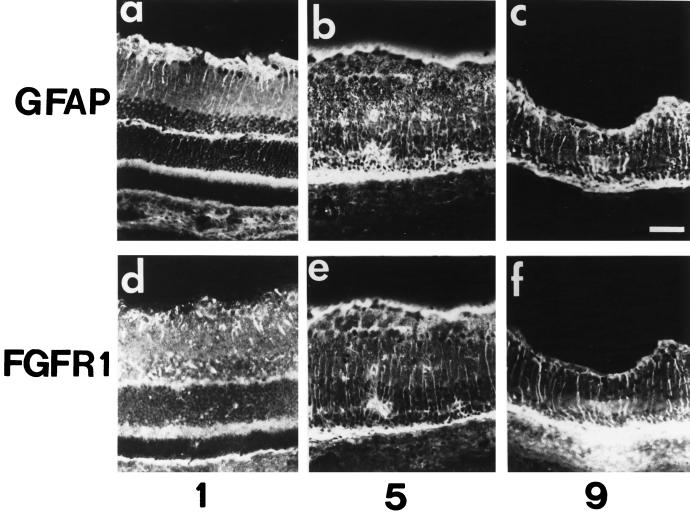
Figure 6.
Immunolocalization of glial fibrillary acidic protein and the full-length FGFR1 in light-induced retina degeneration. Immunohistochemical study of retina of adult rats exposed to constant light for 1 d (1), 5 d (5), and 9 d (9), probed with anti-GFAP antibodies (a–c) and with anti-carboxyl terminal FGFR1 antibodies (d–f), was performed as described in MATERIALS AND METHODS. Note the increase in GFAP immunoreactivity in the inner nuclear and plexiform layers after 5 d of constant illumination in b and c. This staining colocalized with FGF-R1 immunoreactivity (e and f). Bar, 70 μm.

