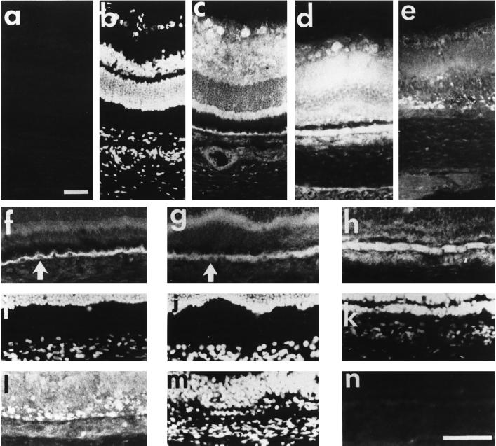
Figure 7.
Immunolocalization of SR1 in light-induced retina degeneration. Immunohistochemical study of retina of nonilluminated control adult rats (a–c, f, and i) and of rats illuminated for 1 d (g and j), 5 d (d, hm and k) and 9 d (e, l, and m), probed with anti-SR1 antibody. Cell nuclei were stained with DAPI (b, i–k, and m). Note the absence of specific staining with the anti-SR1 antibody preadsorbed with the cognate antigen peptide (a and n). (f–n) Nuclear immunolocalization of SR1 in RPE on day 5 of constant illumination; note the absence of SR1 immunostaining in RPE cell nuclei in the control (f and i) and in 1-d–illuminated (g and j) rat (white arrow), whereas in 5-d–illuminated rat (h and k) SR1 was detected in the cytoplasm and in the nucleus of RPE cells. Bars, 55 μm.

