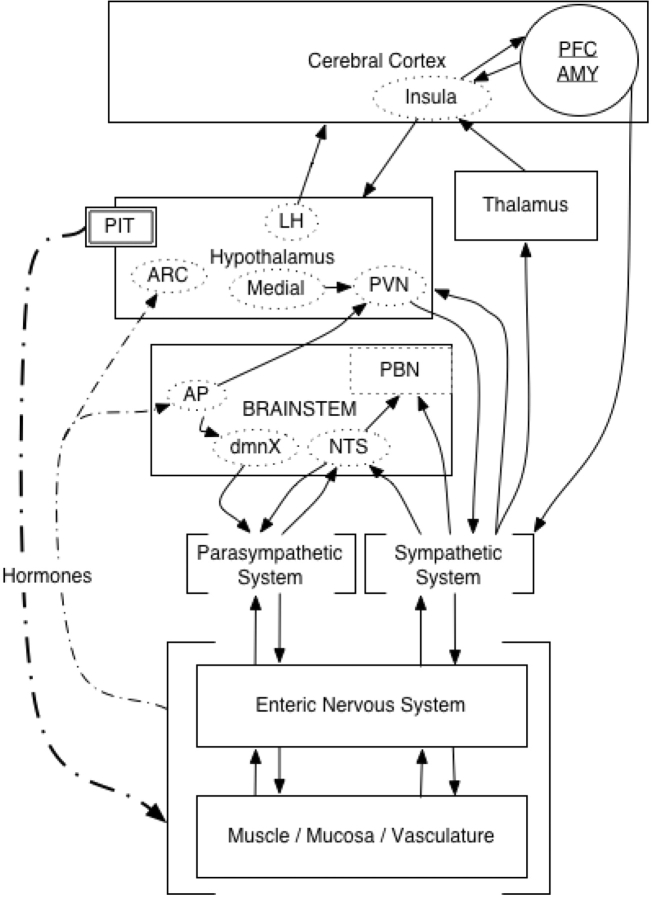
Fig. 1.
Schematic representation of anatomical connectivity between the viscera and central nervous system. Diagram based on Saper (2002) and Craig (2002). Note: AMY, amygdala; AP, area postrema; ARC, arcuate fasciculus; dmnX, dorsal, motor nucleus of vagal nerve; NTS, nucleus tractus solitarius; PBN, parabrachial, nucleus; PFC, prefrontal cortex; PIT, pituitary gland; PVN, paraventricular nucleus.

