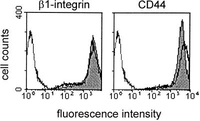
Figure 1.
β1 integrin and CD44 surface expression on MV3 melanoma cells incorporated into 3-D collagen lattices. Cells were cultivated for 14 h in liquid culture (gray area) or within collagen lattice (solid black line), followed by incubation in collagenase and staining with mAbs 4B4 (β1 integrin) or Hermes-1 (CD44) (gray line: isotypic control). Compared with cells cultivated in liquid culture CD44 but not β1 integrin, expression was up-regulated after 14 h of culture in collagen lattices. Additionally, high levels of α3 integrins (fluorescence intensity: 2–6 × 10²) and moderate staining of α5, α6, and αv integrins were detected (0.5–1 × 10²), which is in accordance with previously published data (Klein et al., 1991b; Danen et al., 1993; Goebeler et al., 1996).

