Figure 2.
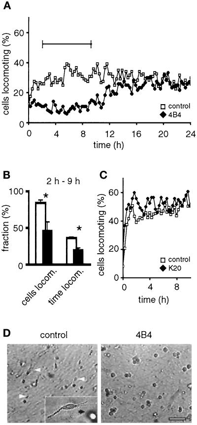
Inhibition of MV3 cell migration in 3-D collagen lattices by adhesion-perturbing anti-β1 integrin mAb 4B4 (A, B, and D), but not by nonblocking mAb K20 (C). Paths of individual cells were digitized, and the percentage of cells locomoting from step to step was obtained. Cumulative percentage of cells locomoting and fraction of time locomoting (B, white bars: control; black bars: mAb 4B4) and representative cell morphologies (D) were obtained for the time period of steady-state locomotion indicated in A (‖—‖). Cells were preincubated with mAb (10 μg/ml) or PBS, washed, and embedded within the collagen matrix. No additional mAb was added to the lattices (A–C), except in D, which contained additional mAb 4B4 in the supernatant. Data represent the mean values (+SD) of three independent experiments (120 cells in total). Asterisks indicate significant differences for P < 0.05. (D) Polarized cell morphology (white arrowheads) was abolished in the presence of mAb 4B4, as detected from video recordings. Black and white cells correspond to cells at different level in depth within the collagen lattice. Bar, 100 μm.

