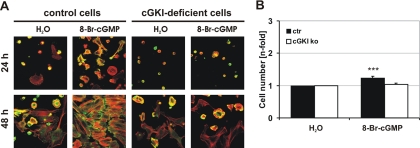
Figure 3.
Analysis of cell adhesion and proliferation. (A) Cytoskeletal staining of primary control and cGKI-deficient VSMCs. Cells were grown on glass coverslips for 24 and 48 h in the absence (H2O) or presence of 8-Br-cGMP (1 mM). VSMCs were stained for F-Actin (red) and vinculin (green) as marker for focal adhesions. Photographs were taken with a confocal microscope (TCS NT; Leica). (B) Primary VSMCs were grown for 72 h under control conditions. Subsequently, 8-Br-cGMP (100 μM) was added for additional 72 h.

