Figure 5.
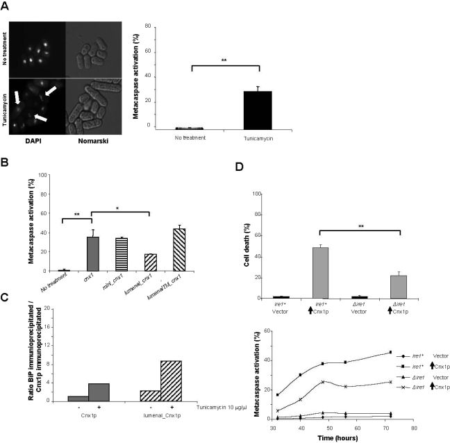
Involvement of calnexin in ER-mediated apoptosis in S. pombe. (A) Tunicamycin induces apoptotic cell death in S. pombe. S. pombe cells carrying a wild-type genomic copy of calnexin (strain SP556) were treated with 5 μg/ml tunicamycin or with the solvent (no treatment) and subsequently stained with DAPI to detect nuclear fragmentation and with FITC-VAD-FMK to determine the % of metacaspase activation. For DAPI staining, cells were examined by fluorescence microscopy. White arrows indicate fragmented nuclei. The % of metacaspase-positive cells was measured by FACS as described in Materials and Methods. (B) Truncation of the TM and the cytosolic tail of calnexin dramatically reduces the levels of metacaspase activation. Cells expressing cnx1+ (strain SP7951R) or mutants of calnexin (mini_cnx1, SP8490R; lumenal_cnx1, SP8488R; and lumenalTM_cnx1, SP8085R) at basal levels were treated with 10 μg/ml tunicamycin, and subsequently the % of metacaspase-positive cells was determined with FITC-VAD-FMK by FACS, as described in Materials and Methods. A global ANOVA showed that the values obtained among the strains were significantly different (p = 0.000). The significance of differences in the results was evaluated by a Student's t test, pairwise calculated with respect to the control for Cnx1p and to Cnx1p for the calnexin mutants. **p < 0.01 and *p < 0.05. (C) Association with BiP. Cells at OD 0.8–1.0 expressing Cnx1p (strain SP7951R) or lumenal_Cnx1p (SP8488R) were treated or not with 10 μg/ml tunicamycin. Immunoprecipitations were performed with anti-Cnx1p antibodies and the membrane was blotted with anti-Cnx1p antibodies or anti-BiP antibodies. Bands corresponding to Cnx1p, lumenal_Cnx1p or BiP were quantified with the Bio-RAD Quantity One 4.6.5 Basic program and reported as a ratio to Cnx1p untreated on a graph. The graph is representative of three different experiments. (D) Implication of Ire1p in apoptosis induced by calnexin overexpression. Cells overexpressing Cnx1p in a wild-type background (SP8007R) or in a Δire1 strain (SP8231R) and the control strain (empty vector, SP7975R and SP8227R) were stained with Phloxin B after 48 h of induction of overexpression to measure cell death. Stained cells were considered as dead. At different time points after the overexpression of Cnx1p cells were stained with the fluorescent probe FITC-VAD-FMK to measure metacaspase activation. Fluorescent cells were quantified by FACS. A global ANOVA showed that the values obtained among the strains were significantly different (p = 0.000). The significance of differences in the results was evaluated by a Student's t test, pairwise calculated with respect to the control for Cnx1p. **p < 0.01 and *p < 0.05. For metacaspase activation, the experiment was repeated three times, and this graph is a representation of a typical experiment. In this figure, vertical black arrows symbolize overexpression of the indicated protein.

