Figure 4.
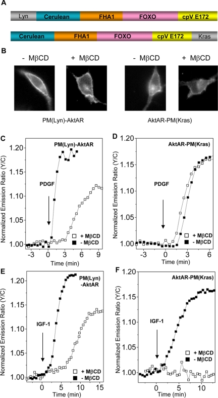
Akt signaling in different microdomains of plasma membrane. (A) Plasma membrane targeted AktAR, PM(Lyn)-AktAR in which the N-terminal portion of Lyn kinase directs AktAR to lipid rafts, and AktAR-PM(Kras) for monitoring Akt activity in the nonraft regions. (B) NIH 3T3 cells overexpressing either PM(Lyn)- AktAR or AktAR-PM(Kras) with or without MβCD treatment. (C) Representative time courses show the responses of PM(Lyn)-AktAR in 50 ng/ml PDGF-stimulated NIH 3T3 cells with (n = 4) or without (n = 3) preincubation with MβCD. (D) PDGF (50 ng/ml) stimulated nonraft Akt activity is not affected with MβCD treatment shown by representative time courses (n = 3). (E) Representative time courses show the responses of PM(Lyn)-AktAR in 400 ng/ml IGF-1 stimulated NIH 3T3 cells with (n = 3) or without (n = 3) preincubation with MβCD. (F) IGF-1 (400 ng/ml) stimulated nonraft Akt activity is abolished by MβCD treatment shown by representative time courses (n = 3∼5).

