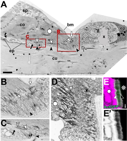
Figure 4.
Ultrastructural features of late larval direct muscle attachments. (A–C) Image of a direct muscle attachment in sagittal view (A) and two close-ups (B and C; as boxed in A). In this preparation, the tendon cell (asterisk) occurs more translucent than surrounding epidermal cells (ep), making its maximal extension (between black curved arrows) easy to visualize. Arrays of microtubules (between black and white arrowheads; open white arrows point at microtubules) are longer toward the distal tip of the myotendinous junction, and the inner layer of the cuticle is bent upward in this area (double chevron in A). Basement membrane reaches to the distal (white arrows in A and B) and proximal (black arrows in A and C) end of the myotendinous junction, but it seems not to penetrate the myotendinous cleft (open black arrows). (D–E′) The cytoskeletal array of another tendon cell showing a relatively broad band of densely coated finger like indentations apically (black arrowhead in D) and the undulating band of the hemi-adherens junction basally (white arrowhead in D); these electron-dense specializations are likely to be the structural equivalents to the apical and basal bands labeled by actin::GFP (ActGFP) or phalloidin in confocal images of tendon cells (arrowheads in E). Bar, 2 μm (A), 0.8 μm (B and C), 1.5 μm (D), and 7.4 μm (E–E′).

