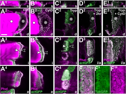
Figure 5.
Properties of F-actin arrays in tendon cells. (A1–E3) Different views of control (con), trypsin-(Tryp), and/or cytochalasin D-treated (CytD) direct muscle attachment sites; planes of view (bottom right) and symbols as explained in Figure 1B. Specimens are labeled with phalloidin (Phal) together with tendon cell-specific actin::GFP (actGFP). The organization of actin into a cytoskeletal belt (between arrowheads in A3) and dashed/stippled fields (A4) clearly resembles tubulin::GFP-labeled specimens (compare Figure 3A). Cytochalasin D destroys most actin in these cells (dots in B2 and E2), but it has little or no effect on the F-actin arrays (B3 and B4), even in orphan tendon cells (E3). Muscle detachment does not significantly affect F-actin arrays in cytoskeletal belt (C3) and stippled fields (C4), and F-actin arrays can still be identified, if detached tendon cells are cultured for another 2 h (D3). (F–F″) Close-up of a detached tendon cell labeled with tubulin::GFP and phalloidin showing the closely intermingled nature of both cytoskeletal fractions. Bar, 10 μm (A–E), 2.8 μm (F′–F″).

