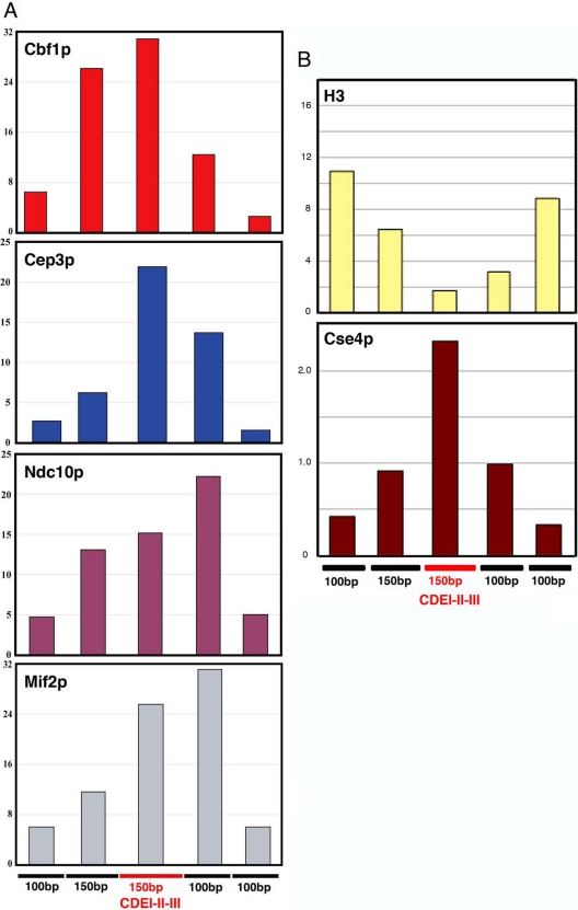
Figure 4.
Chromatin immunoprecipitation of CENIV DNA with Cbf1p-GFP, Cep3p, Ndc10p-myc12, and Mif2p (A) and H3 and Cse4p (B). DNA in immunoprecipitated fractions was amplified by PCR with contiguous, nonoverlapping primers spanning CENIV as shown at the bottom of the figures. Bar graphs show the percentage of IP of each PCR fragment relative to total input DNA.

