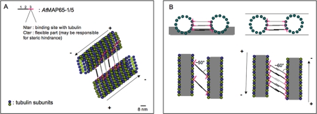
Figure 9.
Schematic representation of AtMAP65-5 or AtMAP65-1 binding along the MT protofilaments. (A) Schematic view of MTs cross-linked by either AtMAP65-5 or AtMAP65-1. AtMAP65-5 or AtMAP65-1 dimerize via domains 1 and 2. The proximal region of domain 3 (red) binds to MTs. The distal region of this domain is unbound and may cause some steric hindrance. The cross-linked MTs are ∼25–30 nm apart. (B) Schematic representation in negative stain and in vitreous ice (top left and right, respectively). Negative stain only partially embeds the cross-linked MTs (side view, above left) and consequently only one level of AtMAP65-1 or AtMAP65-5 is visible in the projected image (below) giving an observed spacing of ∼24 nm. In vitreous ice, the ice specimen is fully embedded (side view, above right). In projection (below) the cross-linking molecules attached to different protofilaments are visible giving the observed repeat of 8 nm. MTs are in gray with outer border represented by tubulin subunits. Scale bar, 8 nm.

