Figure 2.
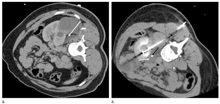
Figures 2, 3. (2) CT after hydrodissection and before RF ablation shows left kidney displaced from psoas muscle with decubitus positioning and percutaneous instillation of 350 mL of 5% dextrose in water (arrow). (3) CT scan during RF ablation shows a left renal mass abutting the psoas muscle after instillation of 5% dextrose in water (white arrow) into the retroperitoneum, with the patient in a near-prone position. The ureter is seen medial to the kidney (black arrow) and is also protected by a fluid pocket.

