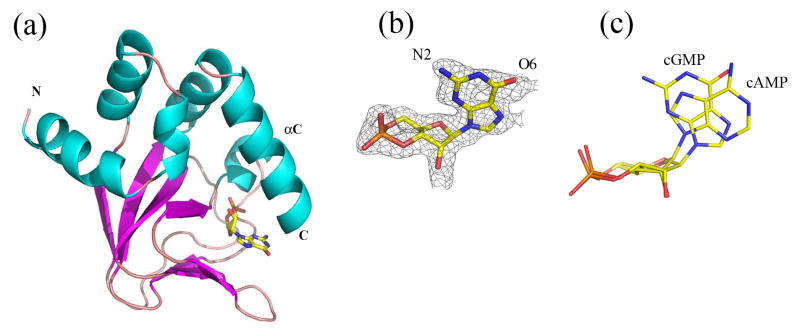
Figure 2. Structure of cGMP bound MlotiK1 CNB domain.
a) The fold of the cGMP-bound wild type CNB domain mirrors that which has been observed for other CNB domains. The ligand sits in a β-roll which is topped by the C-helix. b) Structure and electron density map (simulated annealing omit map contoured at 1.0 sigma) of cGMP bound to the CNB domain. c) Overlay of the ligands from both cAMP and cGMP-bound structures. cAMP is in the anti conformation while cGMP is in the syn.

