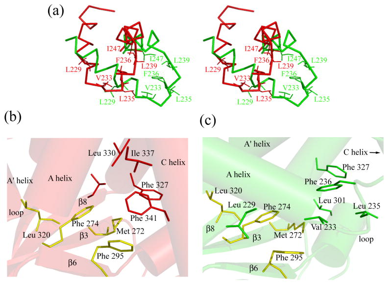
Figure 7. A hydrophobic patch is shielded by the A’- helix/loop/A-helix in the apo structures.
a) Stereo-view of the relative position of the A’-helix/loop/A-helix unit in the bound state (red) and the apo state (green). b) The hydrophobic cluster in the bound state. Residues of the β-roll hydrophobic patch are in yellow. c) The hydrophobic cluster in the apo state. Residues of the β-roll hydrophobic patch are in yellow. The molecules in figures b) and c) were oriented so that residues in yellow superimpose.

